Website invites you to probe a 3-D human brain
Interactive organ offers introduction to neuroscience

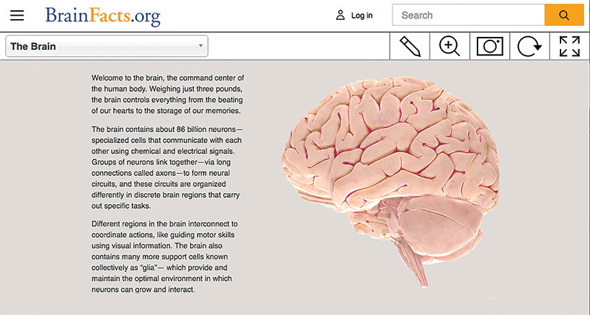
BRAIN TOUR An online, interactive 3-D brain offers an introduction to the organ’s structures.
© Society for Neuroscience 2017
- More than 2 years ago
In movies, exploring the body up close often involves shrinking to microscopic sizes and taking harrowing rides through the blood. Thanks to a new virtual model, you can journey through a three-dimensional brain. No shrink ray required.
The Society for Neuroscience and other organizations have long sponsored the website BrainFacts.org, which has basic information about how the human brain functions. Recently, the site launched an interactive 3-D brain.
A translucent, light pink brain initially rotates in the middle of the screen. With a click of a mouse or a tap of a finger on a mobile device, you can highlight and isolate different parts of the organ. A brief text box then pops up to provide a structure’s name and details about the structure’s function. For instance, the globus pallidus — dual almond-shaped structures deep in the brain — puts a brake on muscle contractions to keep movements smooth.
Some blurbs tell how a structure got its name or how researchers figured out what it does. Scientists, for example, have learned a lot about brain function by studying people who have localized brain damage. But the precuneus, a region in the cerebral cortex along the brain’s midline, isn’t usually damaged by strokes or head injuries, so scientists weren’t sure what the region did. Modern brain-imaging techniques that track blood flow and cell activity indicate the precuneus is involved in imagination, self-consciousness and reflecting on memories.
Clicking and dragging your mouse or finger allows you to rotate the brain. You can zoom in to view areas in detail or back out to get a bigger picture of a region’s connection to the rest of the brain. A drop-down menu provides easy navigation to particular structures and gives more context for the brain’s anatomy. At a glance, the menu outlines, for instance, that the limbic system contains the entorhinal cortex, amygdala and hippocampus. And the hippocampus is further composed of the subiculum and dentate gyrus.
Long, sometimes tongue-twisting names — such as the glossopharyngeal nerve — can be daunting. But the text boxes are easy primers, most no more than 50 to 100 words. Make it through the names and you’ll learn interesting nuggets, like that the glossopharyngeal nerve oversees swallowing muscles and relays information about taste and touch in the mouth.
Anyone interested in the brain should find a trip through this interactive model a fun and informative journey.


