From the May 1, 1937, issue
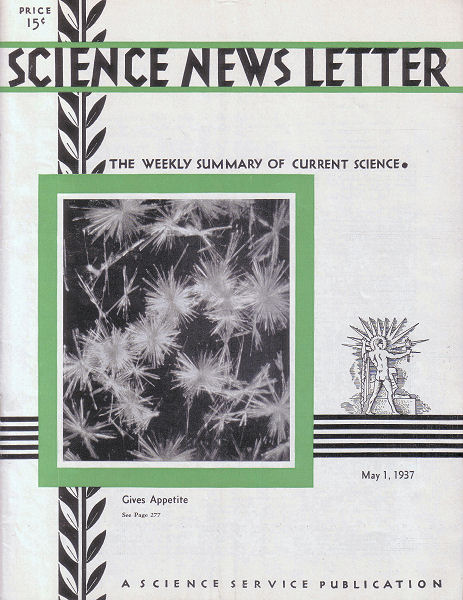
OBTAIN RARE PHOTOGRAPH OF VITAMIN B1 CRYSTALS
Did you ever see a vitamin?
Of course, they don’t really look like the alphabet letters in soup, but did you picture them in your mind as anything like the one shown on the cover of this week’s Science News Letter?
In that rare photograph is shown vitamin B1, the one found in whole cereals, green vegetables, fruit, milk, and yeast, lack of which causes nervous and intestinal malfunctions and loss of appetite and weight.
The picture was taken by Professors W.A. Hynes and Leo Yanowski of Fordham University’s chemistry department. It was taken with a 2-minute exposure by reflected light at a magnification of 20 diameters.
SUGAR IN BLOOD PROTECTS AGAINST POISON OF ALCOHOL
Champion “Old Soaks,” in the days when the West was wild, used to gulp down a lot of olive oil before settling down to an evening of serious drinking. They might have done better had they raided the candy counter instead, it is indicated by recent physiological researches on the effects of alcohol and what to do about them, reported by Drs. Howard W. Haggard and Leon A. Greenberg of Yale University, before the National Academy of Sciences at Washington, D.C.
The intoxicating effect of alcohol on the brain is powerfully counteracted by the amount of sugar in the blood, the two Yale researchers found. The concentration of alcohol needed to kill a rat with one-tenth of 1 percent of sugar in its blood is more than 20 percent higher than the concentration needed to kill when the blood sugar has been reduced to seven-hundredths of 1 percent. But if the blood sugar is raised to two-tenths of 1 percent, the alcohol needed to kill must be increased by 60 percent.
There is a limit to the protective action of sugar, however. Drs. Haggard and Greenberg artificially raised the rats’ blood sugar to concentrations impossible to attain in any natural manner, but found that these did not increase the protective action.
BODY CELLS FOLLOW PATTERN SEEN IN THE BEES’ HONEYCOMB
The bodies of animals and plants are built of cells which are primarily liquid drops. We begin our existence—turning a deaf ear to Aristotelian and scholastic dialectic—as a spherical drop of liquid. We arise as a cell that has the form of a soap bubble or raindrop. The wonderful globular form of a drop of rain is due to an enveloping skin, which has the properties of a stretched elastic membrane. Robert Boyle, in 1676, warily let fall some drops of oil into rectified spirit supernatant to a solution of niter, thinking to explain the structure of the universe. It was the beginning of physical and chemical studies of the tension that abides in the external layer of the drop, whereby the drop constantly strives to contract and occupy the least possible space. It makes a handsome sphere of the egg yolk or the smaller rabbit’s ovum, both of which are single initial cells.
The first drop then divides into a pair, Siamese twinned, joined to one another by a membrane of the same tension as that which covers their exterior. More divisions follow, resulting in a cluster of liquid drops, arranged in accordance with laws established by the Belgian physicist, Plateau, in his masterly study of soapsuds (Statique des Liquides, 1873). Three films, belonging to three bubbles, meet along every edge: six films, belonging to four bubbles, meet at every corner. Hence, as shown by Lord Kelvin, an entire bubble, surrounded by other bubbles of the same size and filling space without interstices, will have 14 facets of contact with its neighbors. If the tension of its walls keeps its surface minimal, it will have eight hexagonal facets and six square facets. Cells of elder pith show a recognizable approach to this ideal pattern. Other cells of plants and animals, more irregular in size and arrangement, likewise present the average of 14 facets, though diverse in outline. There are many pentagonal faces. This outcome is a mathematical necessity for liquid drops in masses when obedient to Plateau’s laws.
When tension causes three facets to meet at every corner of the faceted drop (and any other arrangement is unstable), then the total number of sides of the polygons covering a cell will be 12 less than if they were all hexagons. In a cube, three faces meet at every corner; the six squares which bound the cube have 12 sides less than six hexagons. This will apply to every cell with three-rayed vertices, as, for example, to the 14-hedron with its eight hexagonal and six square faces. It is a corollary of Euler’s famous theorem for all polyhedra, invented by the eminent Swiss mathematician in 1752.
Under these conditions the cellular mosaic, forming, for example, the epidermis of a cucumber, or lining the human intestine, will approach the hexangular pattern of honeycomb. As the cells grow and divide, pentagonal and heptagonal elements are introduced, but the average of six sides is maintained along the tube. Whenever an element is pushed out, regardless of its number of sides, and the gap is closed with none but three-rayed vertices, the mosaic loses just six sides. When, by division, a new cell is added, having any number of sides but making three-rayed vertices only, the mosaic will gain six sides. Under these stringent mathematical requirements, cells present an array of beautiful patterns, complicated by the development of spaces at the corners and edges, where the tension that makes cells round prompts them to separate most readily. For all these patterns there is a simple hydrostatic basis. Cells are fundamentally liquid drops—gland lobules and vascular units are larger drops—all subject to Plateau’s law and to the corollary of Euler’s theorem for polyhedra. Thus neatly, in making cells and glands, “nature Geometrizeth and observeth order.”