Scientists have traced all 54.5 million connections in a fruit fly’s brain
The new ‘connectome’ hints at how information flows
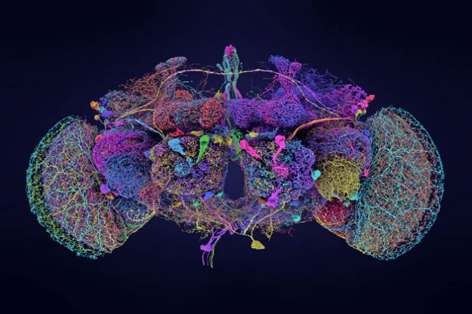
The first full map of fruit fly’s brain charted 139,255 nerve cells and their millions of connections.
Tyler Sloan for FlyWire, Princeton University, (Dorkenwald et al/Nature, 2024)
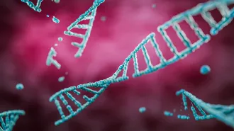
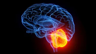
In the brain of a singular fruit fly, nerve cells weave themselves together, enabling flight, mating, eating, sleeping and every other activity of her fly life. Now, in nine papers published October 2 in Nature, scientists report the first complete map of her nerve cells — all 139,255 of them, to be exact — and their 54.5 million connections.
This whole-brain map, traced over years with painstaking precision, is tiny but exquisite: It holds 149.2 meters of neural wiring, all tidily packed into a brain about the size of a poppy seed. As such, this map shows how neural information might flow among cells in Drosophila melanogaster, an animal that’s simpler than a human but complex enough to remain mysterious to people trying to understand its brain.
“This work is absolutely fascinating,” says neuroscientist Olaf Sporns of Indiana University in Bloomington. Back in 2005, he and his colleagues coined the term “connectome,” an accounting of the connections between nerve cells, or neurons (SN: 2/7/14). In the nearly 20 years since then, scientist have mapped more connectomes, including those of male and hermaphrodite C. elegans worms, a larval fruit fly, small bits of mouse and human brains, and part of an adult fruit fly’s brain (SN: 3/9/23; SN: 8/7/19; SN: 5/23/24). This latest fruit fly connectome is the biggest of its sort.
“When connectomics first got started, creating a map like the one presented in this work seemed almost like science fiction,” Sporns says. “And now, amazingly, here it is.”
The project involved electron microscopy images of more than 7,000 thin slices of a female fruit fly’s brain and machine learning that aligned the complex tendrils of neurons, tracing cells through different slices. Machine learning got the researchers within striking distance of the whole connectome. “But humans are still required to correct the errors,” says Sven Dorkenwald, a computational neuroscientist who worked on the project at Princeton University and who is now at the Allen Institute for Brain Science and the University of Washington in Seattle. Hundreds of people from more than 50 laboratories proofread the map with human eyes, ensuring that cells’ shapes were as they seemed to be. It was a big job, from start to finish.
“Did we think it was going to take this long, like, almost 20 years later we would have the fly connectome? Probably not,” says Sebastian Seung, a computational neuroscientist at Princeton University. “But overly optimistic people drive progress.”
In the early days, working on a connectome map “was a contrarian thing to do,” Seung says. “Most people thought it was crazy. There were two objections. One is that it is not possible, and the second is that even if you were successful, the data would be useless.”
But already, the data have proven their utility, revealing cellular details and juicy hints about how brains work. For instance, there are only two CT1 neurons in the whole fly brain, each of which is involved with sensing changes in light and motion. Each neuron stretches across an entire eye and makes a massive number of synapses — more than 148,000, the map shows.
Another analysis sorted some neurons into classes called “integrators,” which receive a huge number of messages from other cells, or “broadcasters,” which send signals to a large audience. These megaphone cells might help signals spread, but in selective ways.
And with the connectome now mapped, scientists have begun to build computer models of how information flows in the brain. “You start with the connections between neurons, and you use that to help you build a simulation of a network,” Seung says. “It’s a totally obvious approach but you couldn’t do it if you didn’t have the connectome.”
One new study, for instance, shows how taste neurons can activate other downstream cells. And that’s just the beginning, Seung says. “My joke for the science fiction enthusiasts is that one fly did have to be sacrificed for this experiment, but this fly could live forever in simulation.”
Sporns also looks to the future: “I foresee a future where connectome maps will become even more comprehensive and detailed, soon to include brains of vertebrates like mouse and human,” he says. Those maps will help answer big questions about brain connectomes — whether they’re variable among individuals, if they change over time, and whether they can help predict behaviors.