The delicate touch of visible light allows scientists to peer into living cells without disrupting them. However, to discern the subtlest details, optical microscopes have long been considered too crude a tool. Instead, researchers have developed techniques such as electron microscopy to make out the finest features, but only in dead specimens.
Enter the stimulated emission depletion microscope. This novel optical device harmlessly resolves fluorescently labeled bits of living cells that are smaller than the so-called diffraction limit, say its developers at the Max Planck Institute for Biophysical Chemistry in Göttingen, Germany. Because light bends, or diffracts, around the edges of objects, ordinary optical microscopes can discern features no closer than a half-wavelength apart, a distance of 200 nanometers (nm) or so.
Beating the diffraction limit, the new prototype instrument can resolve depth to one-sixth of what the best conventional optical microscopes can achieve. Its horizontal resolution comes in under one-half the other instruments’ limit, report Thomas A. Klar and his colleagues in the July 18 Proceedings of the National Academy of Sciences.
Considering all three dimensions, the device can distinguish structures only one-eighteenth the minimum volume discernable by diffraction-limited equipment, the researchers say.
“The work by [the Göttingen team] has the potential to transform the fluorescence microscopy ‘Renaissance’ we are currently experiencing into an ‘Enlightenment Millennium,'” says Shimon Weiss of Lawrence Berkeley (Calif.) National Laboratory in a commentary in the same journal issue.
While the immediate focus is biology, “this beautiful story is not going to end here,” he adds, predicting possible applications in lithography, electro-optics, magneto-optics, and other areas.
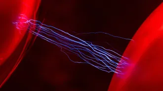
Like other so-called confocal microscopes, the novel instrument works by focusing a laser inside cells tagged with nontoxic fluorescent molecules. By scanning the light across horizontal levels in the cell and then assembling the slices in a computer, researchers can generate a three-dimensional fluorescent image of the cell’s interior.
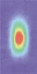
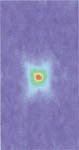
Typically, an oblong laser spot created by a confocal microscope has a diffraction-limited size of about 600 nm in depth and 200 nm in girth, says team leader Stefan W. Hell. What enables the new microscope to both beat the diffraction limit and reduce distortion from its oblong spot, he explains, is a one-two combination of laser pulses.
The device generates a 0.2-picosecond pulse of green light, which creates the typical oblong illumination. Immediately afterward, it fires an oddly shaped and much longer-lasting red pulse into the area excited by the green pulse. The red squelches fluorescence from the green light anywhere the pulses overlap.
Because the second pulse is doughnut-shaped, with blobs of light above and below its doughnut hole, the red light confines fluorescence to a roughly spherical spot 100 nm across. Comparing the green oblong to an American football, Hell says that the final truncated spot resembles a tennis ball. Thanks to that smaller spot size, he adds, the microscope can scan with a finer point and discern unprecedentedly small features.