U.S. and British scientists have devised a gentle way to peer inside living flesh. They use holograms.


The laser-based method eventually may enable doctors to dispense with tissue biopsies on or near external and internal body surfaces, says team leader David D. Nolte of Purdue University in West Lafayette, Ind. Instead, physicians might opt to investigate questionable growths by means of surgery-free, video explorations–”fly-throughs,” as the researchers call them. A system that slices a 3-D hologram into flat images would generate the movie.
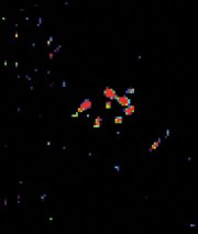
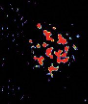
To demonstrate the technique, Nolte and his colleagues grew a rat osteogenic sarcoma–a tumor of bone and connective tissue–in a nutrient broth and then took pictures of it with their new imaging tool.
“This is the first holographic fly-through of living tissue,” Nolte says.
Ping Yu of Purdue will publicly unveil the new method and a video next week in Long Beach, Calif., at an annual conference, known as CLEO/QELS, that’s devoted to lasers, electro-optics, and quantum electronics.
The new approach relies on a novel type of holographic film developed by Nolte’s team. The scientists take advantage of the surprising distance, a few millimeters, that infrared light waves can travel into seemingly opaque tissue.
An ordinary hologram, like those embossed on credit cards, is a laser-made image recorded in plastic as a pattern of fine ridges and grooves. Those topographic features scatter ambient light in such a way that a three-dimensional image appears.
To make such a hologram, a process splits a laser beam into two, bouncing one beam off the object to be imaged, and then reunites the beams. Their interaction generates interference patterns that are recorded in photosensitive materials (SN: 5/22/99, p. 326).
In contrast, Nolte and his coworkers have developed a holographic film–really a semiconductor chip–that redistributes its internal electric fields in response to the split laser beam (SN: 1/8/94, p. 20).
About as thick as a bacterium, the chip is a stack of 200 alternating layers of gallium arsenide and aluminum gallium arsenide. This structure makes the device exquisitely sensitive to light. “It’s the world’s most sensitive holographic film,” Nolte claims.
Using a system of lenses and mirrors, the researchers gather the laser light after it reflects back from inside the tissue. “While most light waves that penetrate the tissue just bounce around wildly inside, a few travel in largely unimpeded, hit some internal structure, and then reflect straight back out again,” Nolte says. Those blend at the chip with unfettered laser light to create a hologram of the tumor interior.
Because the researchers illuminate the tissue with very short laser bursts, they can precisely time the light’s roundtrip travel. It indicates the depth of a burst’s penetration before reflection. Then, by means of a joystick, the researchers control a mirror that permits only light reflected back from a particular tissue depth to contribute to the hologram. As they shift the stick, the researchers can effectively speed through a series of holographic slices. The chip generates a new hologram in less than a millisecond, and the technique can scrutinize slices as thin as 30 micrometers.
“This is a wonderful application of this technique of real-time holography,” comments Gilmore J. Dunning of HRL Laboratories in Malibu, Calif.


