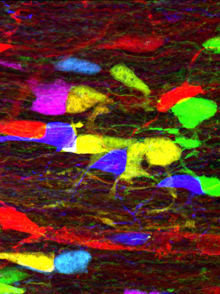
A mouse’s optic nerve glows in a rainbow of colors in the micrograph above.
The image is made using Brainbow, a technique developed in 2007 that inserts genes for fluorescent proteins into animals. When activated, the proteins illuminate some cells in a range of colors.
While most researchers use Brainbow to visualize connections between nerve cells in the brain, Alain Chédotal of the Institut de la Vision in Paris and colleagues customized the technique to trace networks of cells called oligodendrocytes. These cells wrap a material called myelin, the biological equivalent of electrical insulation, around long strands of nerve cells that transmit electrical signals in the brain and throughout the body.
How oligodendrocytes work together to wrap nerve fibers in myelin becomes evident in Brainbow photos of the roughly 3-millimeter-long optic nerve, the team reports in the April Glia. The myelin shields the precious link between brain and eyes.
Studying interactions among oligodendrocytes as well as the cells’ reactions to various drugs may lead to improved therapies for multiple sclerosis, a disease caused by the destruction of myelin.